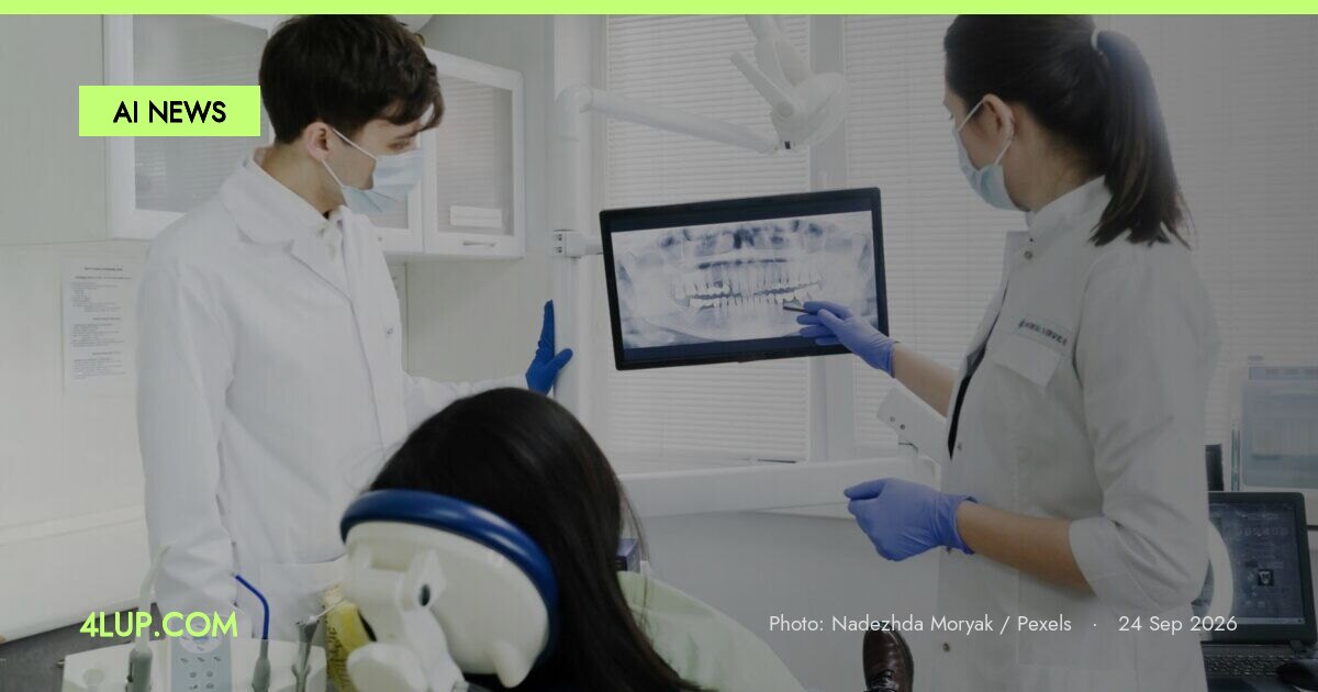
Researchers have introduced a new computational framework designed to overcome the hardware limitations of dental cone-beam computed tomography (CBCT). In many clinical settings, CBCT scanners utilize detector configurations that produce a truncated field of view (FOV), which fails to capture the entirety of a patient’s relevant anatomy. A recent report from the arXiv preprint server details a three-stage method that leverages artificial intelligence to reconstruct the missing anatomical data from these partial scans, effectively extending the viewable area.
The Role of Implicit Neural Representations
The technical approach centers on using implicit neural representations (INR) to fill in the gaps within the truncated projection data. This method represents a shift from traditional voxel-based reconstruction by treating the image as a continuous function. By estimating the missing segments of the scan, the system can then perform an iterative reconstruction to produce a secondary volumetric image. This stage is crucial for ensuring that the generated anatomy remains consistent with the actual physical structures of the patient. The process concludes with a refinement step using a fast diffusion model, a generative AI technique adapted to improve the clarity and accuracy of the final output.
Clinical Implementation and Limitations
The announcement did not specify the specific hardware brands or detector types that were tested with this framework. Additionally, the report does not provide details regarding the processing time required for the diffusion model to complete a reconstruction, which is a critical factor for real-time clinical workflows. It remains unknown how the AI handles complex cases involving dental implants or other dense materials that typically cause streaks and artifacts in CT scans. The research also did not disclose whether the training data included a diverse range of patient ages and jaw structures. Observers should watch for future peer-reviewed studies to determine if this software-based solution can reliably replace the need for larger, more expensive physical detectors in dental offices.
This research demonstrates how generative models can be used to synthesize missing medical data, potentially reducing the need for repeat scans and lowering overall radiation exposure for patients.
Lead image: Nadezhda Moryak / Pexels